Technology That Takes an Up-Close Look
When we’re examining your eyes at Stoney Creek Optometry, diagnostic equipment and technology are vital parts of understanding your eye health during a comprehensive eye exam. Imaging and measuring technology allows us to detect eye diseases or conditions and take an up-close look at significant parts of the eye, such as the retina or cornea.
Request an appointment for a comprehensive eye exam, and our team will guide you through our diagnostic devices.
What to Expect During Diagnostic Testing
When we use diagnostic equipment in our practice, many tests require you to sit facing the machinery with your chin and forehead on a headrest. The imaging process only takes a couple of minutes and is noninvasive.
Our Diagnostic Technology
At Stoney Creek Optometry, we use advanced diagnostic devices to understand your eye health and take a closer look at the eye.
Retinal Imaging
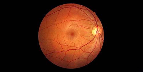
Optical Coherence Tomography is just one specific form of retinal imaging, but there is a wide variety of other imaging techniques, such as fundus photography and angiogram. As with OCT, retinal imaging is used to view the retina in more detail, examine your eye health, and identify any eye diseases or conditions that may not be detected otherwise.
Automated Keratometry
Keratometry is the process of measuring the curvature of the cornea’s surface. Automated keratometry uses light ray reflection and photodetectors to take an image and measure the cornea curve with more precise accuracy.
Automated Keratometry technology is important for assessing the condition of the cornea’s surface, as a damaged corneal surface can lead to vision problems. An automated keratometry test can detect conditions such as astigmatism, where the cornea has an irregular shape that causes blurry vision, and keratoconus, where the cornea appears cone or dome-shaped causing vision issues.
Pachymeter
A Pachymeter is a device that measures the thickness of the cornea to determine if the cornea is swollen and contains excess fluid, which could point to vision problems. A pachymeter is also helpful in determining corneal thickness and health before laser eye surgery to determine candidacy.
Automated Puff Tonometry
Tonometry technology is used to measure the eye’s pressure, also known as the intraocular pressure (IOP). Automated puff tonometry, or non-contact tonometry, measures this using small puffs of air that travel to the cornea.
Measuring the IOP of the eye can help determine if glaucoma might be present, and automated puff tonometry is an important diagnostic tool for glaucoma.
Optical Coherence Tomography (OCT) for Comprehensive Eye Exams
Optical Coherence Tomography (OCT) is a digital imaging technology and the gold standard for examining and detecting diseases of the retina and optic nerve. OCT imaging takes cross-sectional photos of the individual layers of your retina and can create a detailed assessment of your optic nerve.
Using noninvasive light waves, we can precisely map and measure tissue layers to get unique details about your eye tissue, blood vessels, and nerve fibres. This comprehensive information plays a crucial role in the early detection and treatment of several eye diseases.
What Happens During an OCT Scan?
Preparing for an OCT scan is simple—there’s nothing you need to do except focus on a green light.
During the scan, you’ll sit and rest your chin on a support attached to the OCT device. The equipment scans 1 eye at a time. You might see a red line as the scan is completed, but nothing will touch your eye.
The entire process usually only takes a few minutes.
What Conditions Can OCT Help Detect?
OCT is a powerful tool that can help us detect a variety of eye diseases and conditions by precisely mapping intricate details of the eye tissue and vessels at the back of your eye. Moreover, OCT is frequently used to evaluate the optic nerve, a bundle of fibres that carry visual signals from your eye to your brain.
Some of the conditions OCT can assist us in diagnosing include:
- Age-related macular degeneration
- Glaucoma
- Diabetic retinopathy
- Macular edema
- Abnormal blood vessels & blockages
- Cone and cone-rod dystrophies
Is an OCT Scan Covered by OHIP?
Currently, OHIP does not provide coverage for OCT scans. However, some private insurance plans may include coverage for this type of scan. Here at Stoney Creek Optometry, we make it a point to add OCT into every standard comprehensive eye exam.
This helps us personalize your care and gain a detailed understanding of your eye health. By closely assessing your vision and monitoring your eyes for any changes, we can identify potential issues and provide proactive solutions.You can learn more about OCT and OHIP coverage by visiting our blog.

Visit Our Practice Today!
We utilize our technology to provide the best vision care services for you. Visit Stoney Creek Optometry today, and let’s take a look at your eye health.


Our Brands


Extended Hours, Direct Billing, & Conveniently Located
Life can get busy, and we want to help simplify your eye care experience. We are conveniently located on all major bus routes, provide free parking, have extended evening and Saturday hours, and offer direct billing to most insurance companies. We implement our extensive experience and innovative technology into our eye exams to ensure your visit is a productive one.

Our Location
Our clinic is located on Queenston Road, right next to Fortinos Plaza in the prestigious Stoney Creek Professional Arts Building.
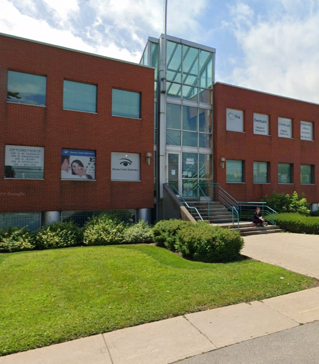
Our Address
- Stoney Creek Professional Arts Building, 980 Queenston Rd. Suite 202
- Stoney Creek, ON L8G 1B9
Contact Information
- Phone: 905-664-5949
- Email: [email protected]
Hours of Operation
- Monday: 8:00 AM – 5:00 PM
- Tuesday: 8:00 AM – 7:00 PM
- Wednesday: 8:00 AM – 5:00 PM
- Thursday: 8:00 AM – 7:00 PM
- Friday: 9:00 AM – 5:00 PM
- Saturday: 9:00 AM – 1:00 PM
- Sunday: Closed

Our Google Reviews